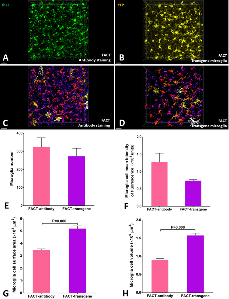
Figure 8.
The FACT protocol is compatible with immunohistochemistry and performs well in both antibody-based and transgene-based imaging. (A and B) Three-dimensional 200-µm-thick volumes of cerebral cortex after antibody staining and YFP-labeled microglia in transgenic mice. (C and D) Three dimensional reconstructions of microglia surface area and volume by the Imaris Surface algorithm and color sorting of the areas by Imaris Vantage from white (highest area) to purple (lowest area). Higher detection of microglia signal was achieved in the Iba1 immunostaining compared to transgene-YFP-microglia . (E and F) Comparisons of corrected microglia numbers and corrected mean signal intensities between the two FACT-based detection protocols after semiautomatic counting with the Imaris Surface algorithm. No significant differences were evident (p > 0.05). (G and H) Comparisons of microglial cell surface area and microglial cell volume between the two FACT-based detection protocols after semiautomatic measurement with the Imaris Surface algorithm. Greater signal detection was seen in the antibody-labeledmicroglia compared to the transgene-labeled microglia (p < 0.05). Scale bars are 100 μm.

