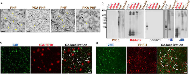
Figure 3.
Electron Microscopy of paired helical filaments (PHF) and PKA treated PHF, and comparative detection of specific NDD conformers. (a) EM of purified PHF from a human AD brain and protein kinase A treated PHF. Yellow arrows show fibrils and black arrows show oligomers in different aggregation clusters. Left two panels show overall difference of fibrils and oligomers in both samples and right two panels are higher magnification to show the oligomers associated to fibrils or in independent clusters. All scale bars are 50 µm. (b) Immunoblots of recombinant deer PrP (grey dPrP); Aβ1–42 freshly dissolved, fibrilized or polymerized (red Aβ42, Aβ42f and Aβ42p respectively), and PHF and PKA PHF (brown). Individual specificity of commercial antibodies PHF-1 for hyperphosphorylated tau; 4G8 and 6E10 for Aβ peptides and 7D9 and 6D11 for PrP protein is compared to the cross-reactivity of hybridomas 10E and 23B (blue) reactive to more than one conformer and oligomeric forms (the uncropped blots showing 10E and 23B reactivity are shown in Supplemental Figure 6). (c and d) Co-localization of hybridoma 23B with either 4G8/6E10 or PHF-1 antibodies on human AD brain tissue. Scale bar represents 100 µm.

