
Figure 1.
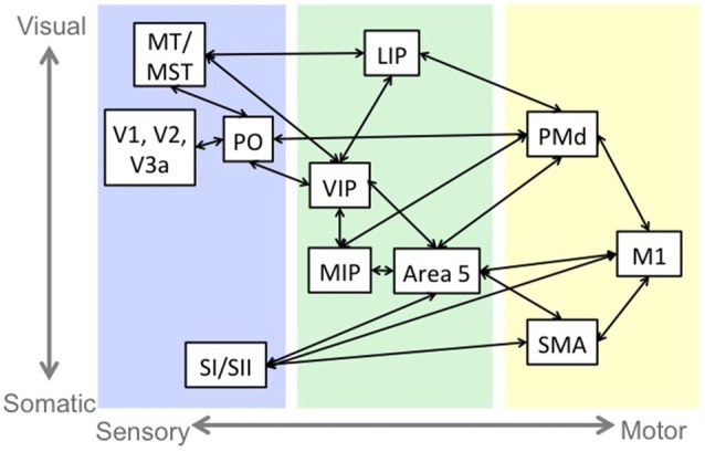
An example of sensorimotor circuitry: the reach pathway. Blue indicates regions that are primarily responsive to sensory stimulation. Green indicates regions that have sensory and motor responses, and yellow indicates regions that have primarily motor related activity. There is a tendency in the sensory and sensorimotor areas for regions near the top of the figure to represent visual information and the regions near the bottom to represent somatosensory information. Abbreviations: middle temporal area (MT), medial superior temporal area (MST), primary visual cortex (V1), secondary visual cortex (V2), visual area 3a (V3a), parieto-occipital area (PO), primary somatosensory cortex (SI), secondary somatosensory cortex (SII), lateral intraparietal cortex (LIP), medial intraparietal cortex (MIP), ventral intraparietal cortex (VIP), dorsal premotor cortex (PMd), primary motor cortex (M1), supplementary motor area (SMA).