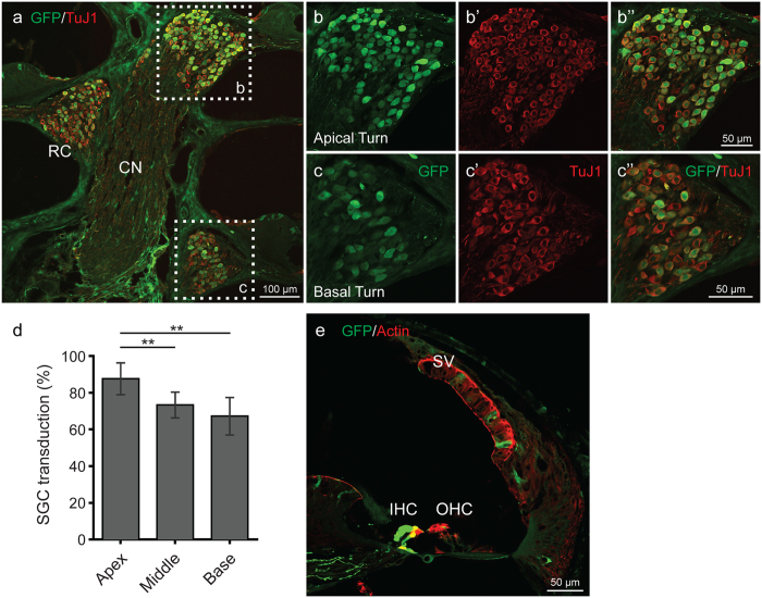
Figure 3.
rAAV2/9 transduction in the spiral ganglion cells (SGCs) and organ of Corti 4 weeks after virus inoculation at P0–1. (a) Midmodiolar cross-sectional images show transduced SGCs in Rosenthal’s canal (RC) (CN, cochlear nerve). Samples were immunostained with anti-GFP (green) antibody and anti-TuJ1 (red) to label SGCs. (b,c”) Images in cross-sections of the apical (b–b”) and basal (c–c”) turns are high-magnification views of the regions marked with white dotted squares. (d) Percent of transduced SGCs in RC. Data are means ± SD from four sections in each of two ears. eGFP expression of SGCs in RC was greater in the apical turn than in the basal turn (*p < 0.05; **p < 0.005). (e) In a cross section confocal image, rAAV2/9 transduced some cells in the stria vascularis (SV). Images were immunostained with anti-GFP (green) antibody and phalloidin (red) to label filamentous actin.

