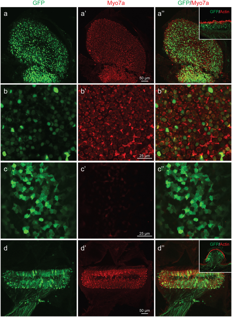
Figure 4.
rAAV2/9 transduction in the vestibule 4 weeks after virus inoculation at P0–1. All images, except insets, were stained with Myo7a (red) for labeling hair cells (HCs) and imaged for native eGFP (green). (a–a”) Confocal images of whole mounts of the utricle. eGFP expression is evident throughout the utricle. (b–b”) High-magnification images of the HC layer show transduced HCs (eGFP-positive cells). (c–c”) High magnification images of the SC layer transduced supporting cells (eGFP-positive cells). (d–d”) Confocal images of whole mounts of the crista ampullaris (CA). eGFP expression is evident throughout the CA. Insets: Cross-sectional images of the utricle and CA. Images were stained with eGFP (green) and phalloidin (red) for labelling hair cells and filamentous actin, respectively. Note hair cell and supporting cell transduction consistent with whole mounts figures.

