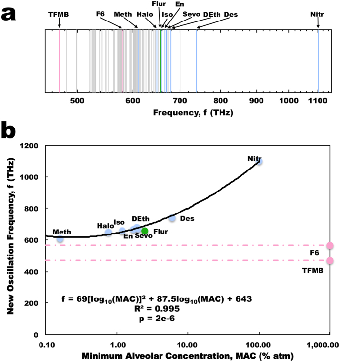
Figure 3.
Collective dipole modes of oscillation in tubulin. (a) Average energies of the collective dipole modes of oscillation in tubulin. Gray – normal modes predicted for tryptophan, tyrosine and phenylalanine in tubulin in the absence of agents. (Blue – additional normal modes introduced due to the presence of an anesthetic agent; Red - additional normal modes introduced due to the presence of a non-anesthetic agent; Green – additional normal mode introduced to the presence of the anesthetic/convulsant agent flurothyl). (b) Agent-induced new frequency modes of oscillation versus MAC. As the non-anesthetics fall below the trend line minimum there is no predicted MAC for non-anesthetics available at any value. (Blue points – anesthetics; Red points - non-anesthetics; Green points – anesthetic/convulsant; Red line – difference between non-anesthetic predicted and actual (~1000% atm) MAC).

