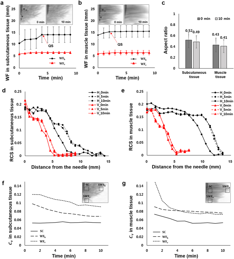
Figure 6.
Temporal variations of WFh and WFv in the (a) subcutaneous tissue and (b) muscle tissue, respectively. Insets are X-ray images of the subcutaneous and muscle tissues at 0 min and 10 min after drug injection. (c) Aspect ratios in the subcutaneous and muscle tissues at 0 min and 10 min after drug injection. Variations of RCS values according to distance from the needle in (d) the subcutaneous tissue and (e) the muscle tissue. Temporal variations of Cv in the (f) subcutaneous tissue and (g) muscle tissue, respectively. The Cv values are measured at the regions of SC, SWFh, and SWFv (insets, dotted boxes). Scale bar: 500 μm.

