Figure 5.
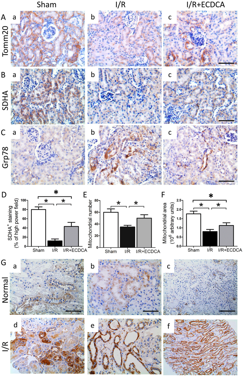
FXR activation protects mitochondrial function and structure and inhibits ER stress during I/R-induced injury. (A–C) Representative images showing immunostaining for (A) Tomm20, (B) SDHA and (C) Grp78 on renal sections from (a) sham, (b) I/R and (c) I/R + ECDCA groups (scale bar 50 μm). Note all three protein markers are localized in proximal tubules. (D–F) Quantitative analysis of (D) SDHA staining per high power field, quantitative analysis of (E) mitochondrial number and (F) mitochondrial area. n = 6 mice/group. Data are means ± SEM, one-way ANOVA with Bonferroni’s test. *p < 0.05. (G) Markers for oxidative and ER stress in normal and preimplantation human renal biopsy specimens. Shown are representative images of immunostaining for (a and d) 4-HNE, and (b and e) GRP78 in kidney specimens obtained from (a and b) normal biopsies and (d and e) preimplantation biopsies (magnification 400x). (c and f) CHOP in kidney specimens obtained from (c) normal biopsies and (f) preimplantation biopsies (scale bar 100 μm).

