Figure 7.

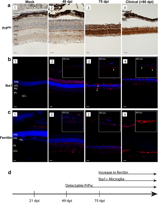
Distribution of PrPSc, Iba1, and ferritin in hamster retinas. (a) Immunoreaction for PrPSc shows no reactivity in mock-treated control retinas (panel 1). PrPSc deposits are first evident at 49 dpi, and are localized to outer segments of photoreceptor cells, OPL, INL, IPL, and GCL (panel 2). Robust PrPSc deposits are seen throughout the retina at 75 dpi and in clinical samples (panels 3 & 4). (b) Immunoreaction for Iba1 shows minimal reactivity in RPE, OPL, and IPL in mock treated samples (panel 1). Prominent reactivity for Iba1 is first seen at 49 dpi (panel 2). Amoeboid microglia are evident throughout the retina, including the RPE cell layer in scrapie-infected samples (panels 3 & 4). Arrows indicating large cell bodies of activated, Iba1 + microglia at later time points (panels 3 & 4). (c) Immunoreaction for ferritin shows minimal reaction in mock-treated samples (panel 1). Increased perinuclear and cytoplasmic immunoreactivity for ferritin is first evident at 49dpi, and is localized to RPE, OPL, INL, IPL, and GCL (panels 2–4). High magnification (60X) inserts in the upper right hand corner show Iba1 and ferritin immunoreactivity in the RPE cell layer in scrapie-infected samples. Abbreviations: GCL: ganglion cell layer; IPL: inner plexiform layer; INL: inner nuclear layer; OPL: outer plexiform layer; ONL: outer nuclear layer; RPE: Retinal pigment epithelium. Scale bars: 10 μm. (d) The increase in ferritin reactivity occurs subsequent to PrPSc accumulation, and coincides with Iba1 reactivity.
