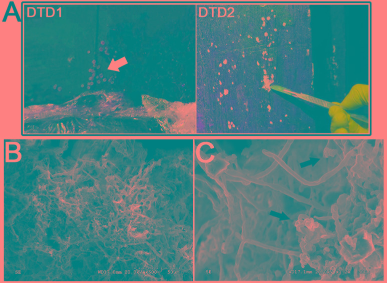
FIGURE 2.
Morphological observations of the heavy fungal colonization of the wooden tomb. (A) Sample DTD1, which exhibited a sporocarp appearance was used for microbial isolation and identification. Sample DTD2 was collected from white spots on the surface of the tomb. (B,C) Scanning electron micrograph of a white spot. Visualization of fungal hyphae and spores which appear ellipsoidal and spherical. The small dots indicate the bar.

