
Figure 1.
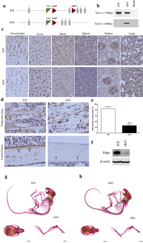
(a) Schematic of the Pdpn floxed allele before and after deletion of the loxP cassette containing exon 3 via osteocalcin cre (Oc‐cre) mediated recombination. (b) PCR analysis of genomic DNA from the long bones of fl/fl, and cKO mice with primers for the Tm1c allele (before cre recombination), and the Tm1d allele (∼440 bp, after cre recombination). (c) Immunohistochemical labeling of Pdpn in the lung, kidney, spleen, heart, liver, muscle, and growth plate, of 6‐week‐old mice. Images are representative of n = 4/sex/genotype. Scale bar = 20 μm. (d) Immunohistochemical labeling of Pdpn in the trabecular bone and cortical bone of 6‐week‐old mice. Arrows are pointing at embedded osteocytes within the trabecular and cortical bone and their dendritic processes projecting from the cell bodies. Images are representative of n = 4/sex/genotype. Scale bar = 20μm. (e) Quantification of osteocytes positive for Pdpn immunolabeling relative to negatively labeled osteocytes (n = 3/genotype), p < 0.001***. (f) Western blotting for Pdpn (∼37 kDa) in cortical bone protein lysates from 6‐week‐old mice. β‐actin was used as a loading control. Whole mount Alcian Blue and Alizarin Red stained skeletal preparations of 6‐week‐old male (g) fl/fl, and (h) cKO mice (scale bar = 10 mm) including hindlimb and calvaria preparations (scale bar = 5 mm)