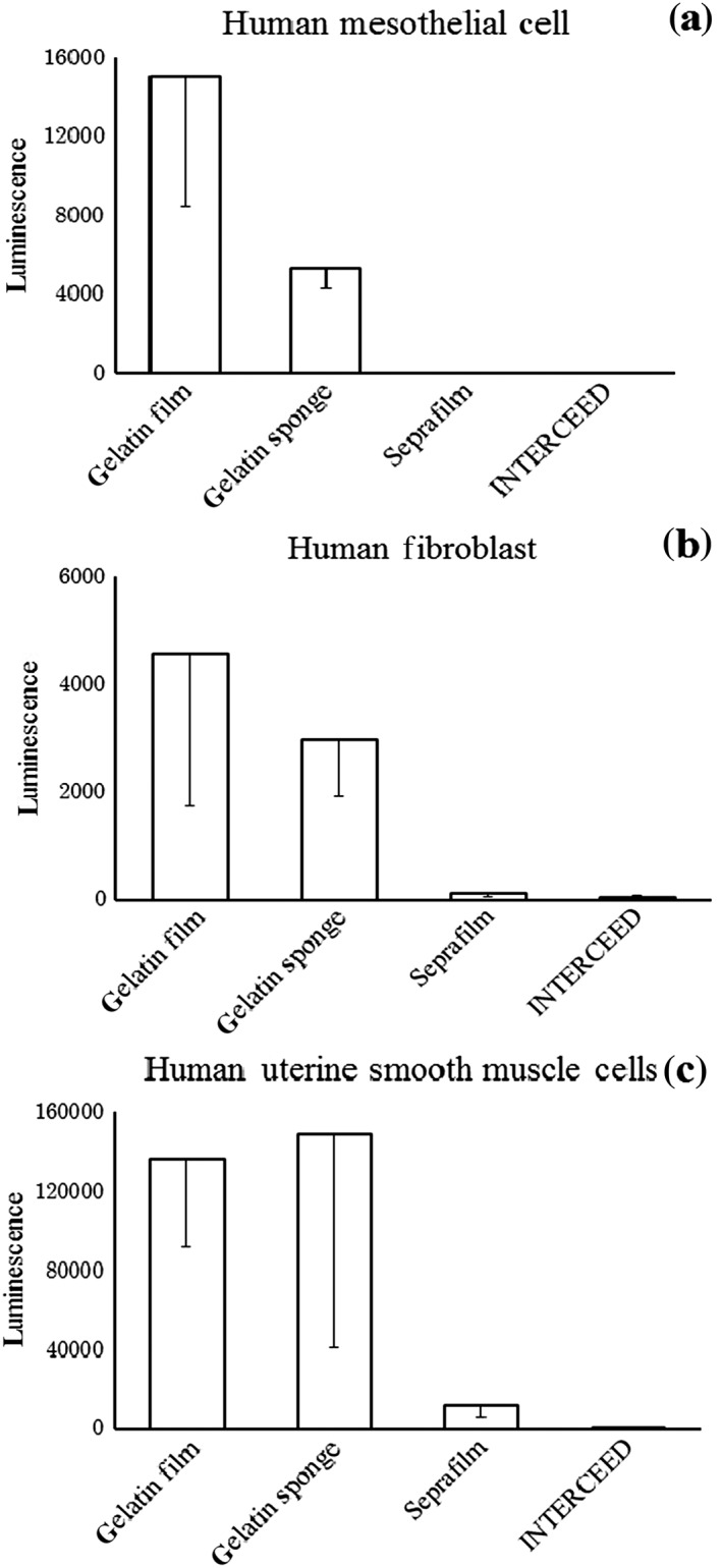
Figure 4.

Cell growth on the materials (1 week): (a) human mesothelial cells, (b) humane fibroblasts and (c) human uterine smooth muscle cells. Significantly richer cell growth was observed in the gelatin film and sponge groups, which corresponded to the both surfaces of the two‐layered gelatin sheet, (P < 0.01) than in the Seprafilm or INTERCEED groups.
