Figure 1.

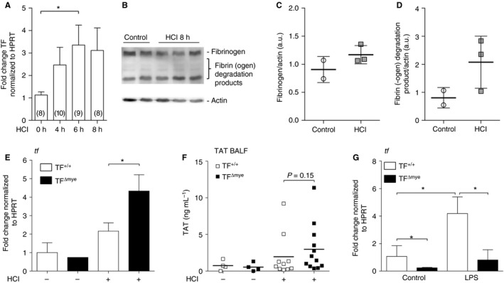
Tissue factor (TF) expression and activation of coagulation during acute lung injury (ALI). (A) TF mRNA expression in lungs of wild‐type mice after HCl‐induced ALI was determined by qPCR at the indicated time points. Data show fold control (0 h) values normalized to hypoxanthine guanine phosphoribosyl transferase (HPRT); numbers are shown in parentheses. (B) Western blot analysis for fibrin‐β of homogenized lung samples 8 h after HCl treatment (n = 3) and of controls (n = 2); actin was used as a loading control. (C, D) Western blot densitometric analysis of (C) fibrinogen and (D) the major fibrin(ogen) degradation product. (E) TF mRNA expression in total lung tissue of control (n TF +/+ = 2; n TF Δmye = 1) and HCl‐treated (n TF +/+ = 9; n TF Δmye = 4) myeloid TF‐deficient (TF Δmye) or wild‐type (TF +/+) mice was analyzed by q‐PCR 8 h after treatment. (F) Thrombin–antithrombin (TAT) complexes in the bronchoalveolar lavage fluid (BALF) of control (n = 4) and HCl‐treated (n TF +/+ = 10; n TF Δmye = 12) wild‐type or myeloid TF‐deficient mice 8 h after treatment. (G) TF mRNA expression of alveolar macrophages, isolated from lungs of myeloid TF knockout mice and wild‐type littermates and stimulated with lipopolysaccharide (LPS) (100 ng mL−1) in vitro for 3 h; n = 5. For statistical analysis, one‐way‐anova with Dunn's multiple comparisons test (A), unpaired Student's t‐tests (E, F) or the Mann–Whitney test (G) were performed; *P < 0.05. Littermate‐controlled experiments were performed. a.u., arbitrary units.
