
Figure 1.
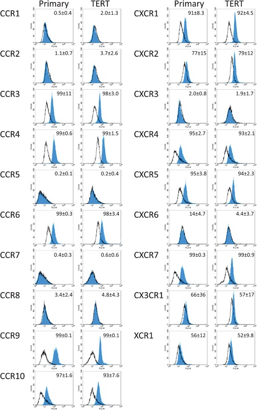
Chemokine receptor expression on gingiva fibroblasts. The expression of chemokine receptors on primary (left column) and hTERT‐immortalized (right column) human gingiva fibroblasts was analyzed by flow cytometry. Open lines represent isotype controls and closed lines the chemokine receptor specific antibody. Mean of percent positive cells and standard deviation of three individual experiments are shown in the upper right corner of each histogram. A total of 12 of the 19 chemokine receptors were expressed