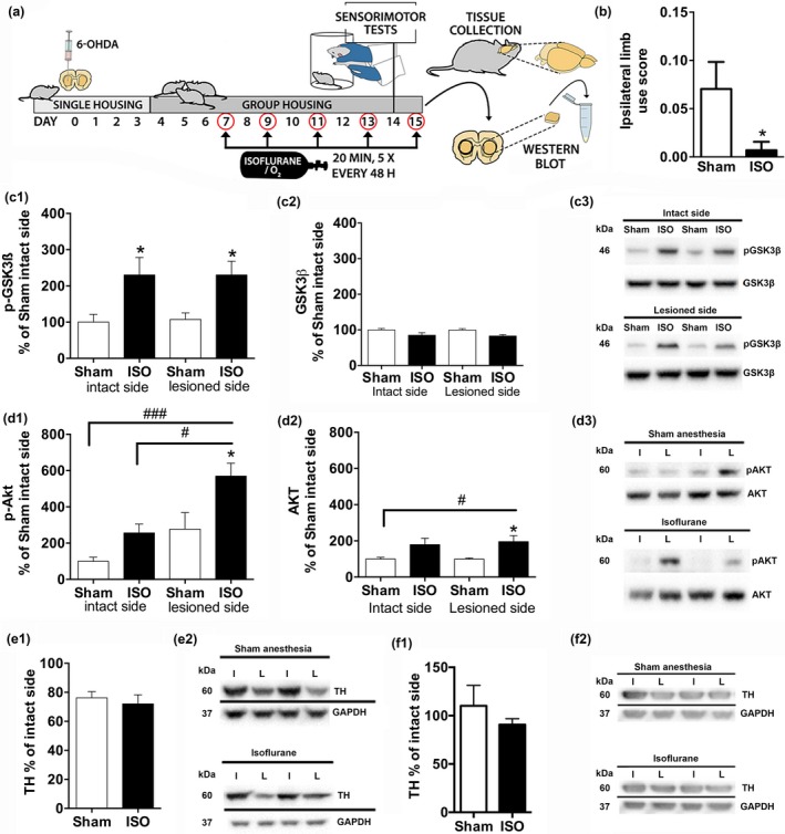
Figure 2.

Isoflurane anesthesia exposure regulates striatal AKT‐GSK3β signaling and ameliorates motor deficits in an animal model of early‐stage Parkinson′s disease. (a) Experimental workflow. Unilateral intrastriatal 6‐hydroxydopamine (10 μg) administration was performed on day 0. Animals were exposed to isoflurane (4% induction for 2 min, 2% maintenance for 20 min; N = 9) or sham conditions (rats in the induction chamber for 2 min with O2 flow on; N = 9) on days 7, 9, 11, and 13. On day 14 (24 h after previous anesthesia exposure), animals were subjected to behavioral tests, assessing sensorimotor functions (cylinder, vibrissae, movement initiation, and adjusting step tests). The animals were exposed to an additional isoflurane/sham anesthesia treatment on day 15, after which the striata were collected for analyses. (b) Effect of repeated isoflurane anesthesia exposure on motor functions (combined ipsilateral limb use score of sensorimotor performance); (c) Phospho‐GSK3βSer9/total‐GSK3β ratio and total GSK3β normalized to GAPDH; (d) Phospho‐AKTThr308/total‐AKT ratio and total AKT normalized to GAPDH; (e) The ratio of TH protein in lesioned versus intact striatum. (f) The ratio of TH protein in lesioned versus intact substantia nigra area. #/*p < 0.05; ###/***p < 0.005, Student t‐test (b, e–f) or two‐way anova followed by Newmann–Keuls post hoc test (c–d). GAPDH, Glyceraldehyde 3‐phosphate dehydrogenase; GSK3β, glycogen synthase 3β; AKT, protein kinase B; ISO, isoflurane; I, intact hemisphere; L, lesioned hemisphere; TH, tyrosine hydroxylase.
