Figure 4.

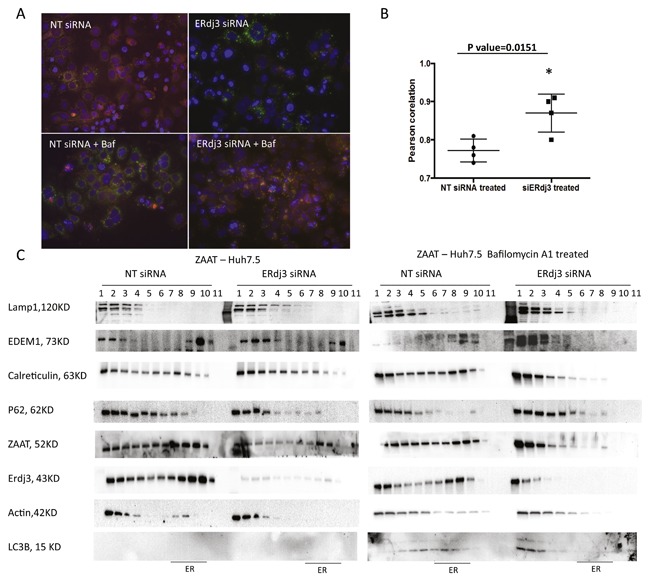
Increased autophagic response to ZAAT accumulation mediated by ERdj3 depletion. (A) LC3‐positive autophagosomes co‐localize with ZAAT in presence of bafilomycin, upon siERdj3 treatment. NT siRNA or 20 µM of siERdj3 was introduced 24 h post ZAAT transfection to AAT KO Huh 7.5 cells. Then, 48 h later, the cells were treated with or without bafilomycin for 6 h. ZAAT was immunostained using mouse anti‐AAT antibody (Alexa 488; red) and rabbit anti‐LC3 (Alexa 594; green). (B) Pearson correlation coefficients graph from four different images with siERdj3‐ versus NT siRNA‐treated samples. (C) Density gradient isolation of cellular proteins from the ZAAT‐expressing cells treated with NT siRNA or siERdj3 in presence and absence of Bafilomycin A1 shows that siERdj3 activates autophagic response to ZAAT accumulation. siERdj3 and bafilomycin A1 treatment results in ZAAT appearance in light‐density fraction, accompanied by EDEM1 and calreticulin in LC3‐ and Lamp1‐positive compartments.
