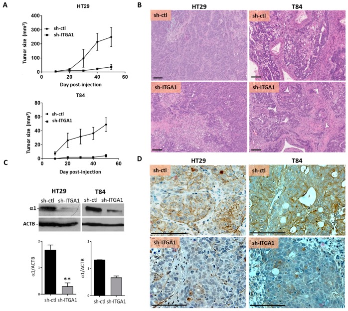
Figure 5.
Integrin α1 subunit/ITGA1 knockdown reduced the development of colorectal tumors in xenografts. (A) Tumor growth assessment after injection of 2 × 10⁶ HT29 or T84 cells into the subcutaneous tissue of nu/nu mice. For both cell lines, the sh-ctl and sh-ITGA1 cells were injected after 10 days of selection with puromycin (10 µg/mL). Tumors were measured in two axes and the volume was determined using the formula V = (D × d2)/2. (B) Representative micrographs of the histological architecture of the tumors derived from sh-ITGA1 and sh-ctl cells for HT29 and T84 cell lines. Hematoxylin and eosin staining (H&E). (C,D) Representative Western blot and immunohistochemical micrographs showing the validation of the repression of expression of the α1 subunit, after resection of tumors derived from sh-ctl and sh-ITGA1 cells, for HT29 and T84 cell lines. N = 3. Student’s t test. * p < 0.05, ** p < 0.01, *** p < 0.001. Scale bar = 100 µm.

