Figure 1.
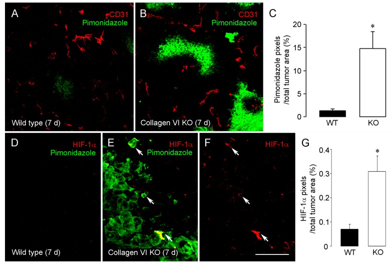
Increased hypoxia and HIF1α expression in tumors in collagen VI null mice. Hypoxia levels in 7-day intracranial B16F10 tumors were determined after intravenous injection of a pimonidazole hypoxia probe (60 mg/kg, 1 h circulation period). Tumor sections were double-stained for pimonidazole (green) and CD31 (red). In contrast to rare pimonidazole-positive regions in wild type tumors (A), areas of intratumoral hypoxia were markedly increased in tumors from collagen VI null mice (B). Intratumoral hypoxia levels, defined as the percentage of total tumor area covered by pimonidazole pixels, were increased 10-fold in collagen VI null mice (C). Very low levels of immunolabeling for HIF-1α (red) were detected in tumors in wild type mice (D), in agreement with the virtual absence of immunolabeling for pimonidazole (green). In tumors in collagen VI null mice, increased hypoxia detected via pimonidazole labeling was accompanied by more abundant labeling for HIF-1α ((E) and (F)). HIF-1α levels are more than 3-fold higher in collagen VI null mice than in wild types (G) * p < 0.05 vs. wild type. Scale bar: 120 μm in (A) and (B); 40 μm in (D–F). Data taken from (You et al. [24]).

