Figure 3.
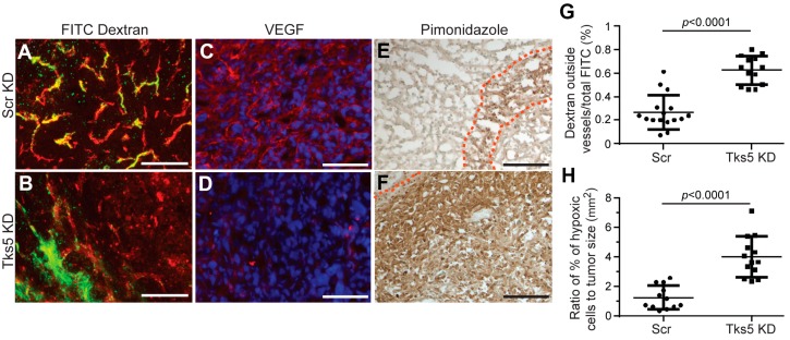
Reduced Tks5 expression in mammary tumor cells results alters tumor blood vessels. Blood vessels in mammary tumors formed by MDA-MB-231 cells transfected with scrambled shRNA (Scr KD) and Tks5 shRNA (Tks5 KD) were analyzed for leakage of FITC-dextran, localization of VEGF-A, and levels of tumor hypoxia. (A) and (B): Levels of FITC-dextran (green) outside of CD31-positive vessels (red) were determined by confocal microscopy. Increased leakage from vessels in Tks5 knockdown tumors is quantified in (G). (C) and (D): Confocal microscopy of immunolabeling sections was used to localize VEGF-A (red) to tumor vessels in control tumors. This vascular localization is largely lost in Tks5 knockdown tumors. Blue = DAPI. (E) and (F): Pimonidazole was injected intravenously into tumor-bearing mice and allowed to circulate for 10 min. Immunolabeling was then used to localize and quantify pimonidazole accumulation in areas of hypoxia. Dashed lines delineate the borders of hypoxic areas. Hypoxia is greatly increased in Tks5 knockdown tumors (H). Scale bars: 100 μm. Data taken from Blouw et al. [29].

