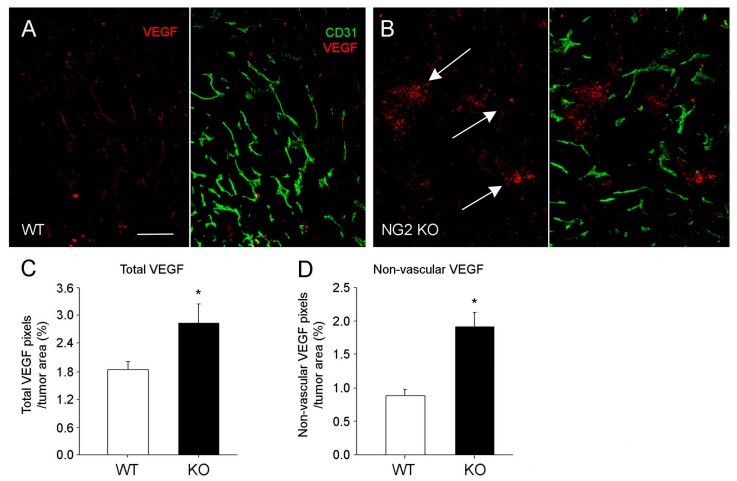
Figure 4.
Localization of VEGF-A expression in mammary tumors. Double immunolabeling for CD31 (green) and VEGF-A (red) was used to localize VEGF-A expression relative to mammary tumor blood vessels in control (A) and NG2 null (B) mice. Total VEGF-A is elevated in tumors in NG2 null mice (C), but this increase is due to an increase in non-vascular VEGF-A (D). In tumors in control mice, VEGF-A expression is largely associated with the vasculature, while more diffusely localized VEGF-A is evident in tumors in NG2 null mice (arrows in B). * p < 0.01. Scale bar: 60 μm. Data taken from Gibby, et al. [31].

