Figure 4.
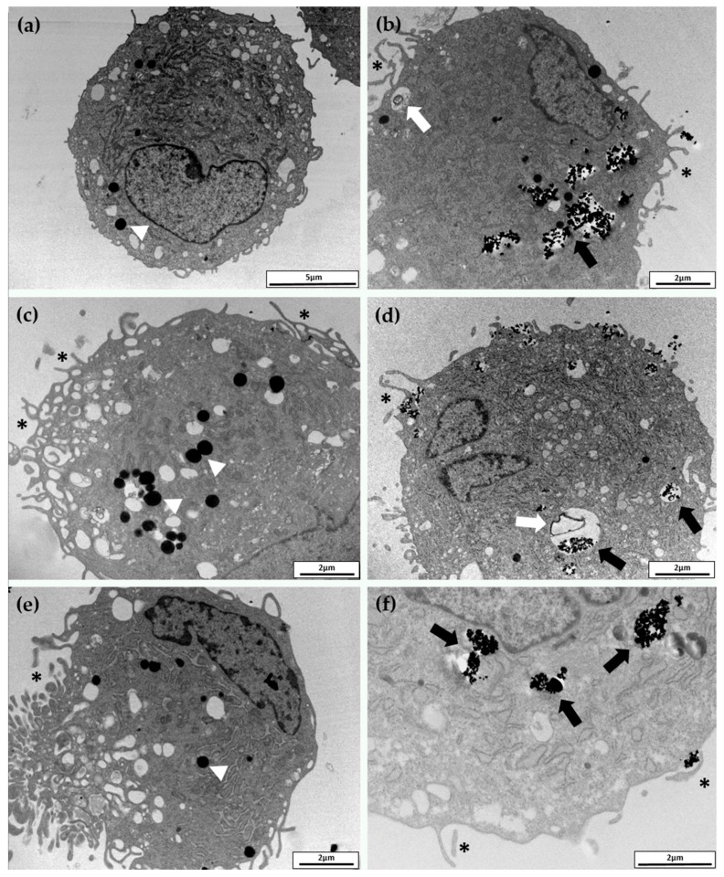
Transmission electron microscopy images of typical hASCs exposed for 24 h to FeMPs (a), FeNPs (b), CoMPs (c), CoNPs (d), NiMPs (e), and NiNPs (f). NPs were identified as high-electron-density objects when inside the cell and localized inside the vesicles (black arrows). No differences were observed among the three considered NPs and the internalization appeared to be nonspecific. White arrows indicate lysosomes of different sizes containing amorphous material. Several pronounced pseudopodia-like protrusions (*) are present both in NP- and in MP-exposed cells. Nuclei and mitochondria do not contain NPs. Arrowheads indicate lipid droplets.

