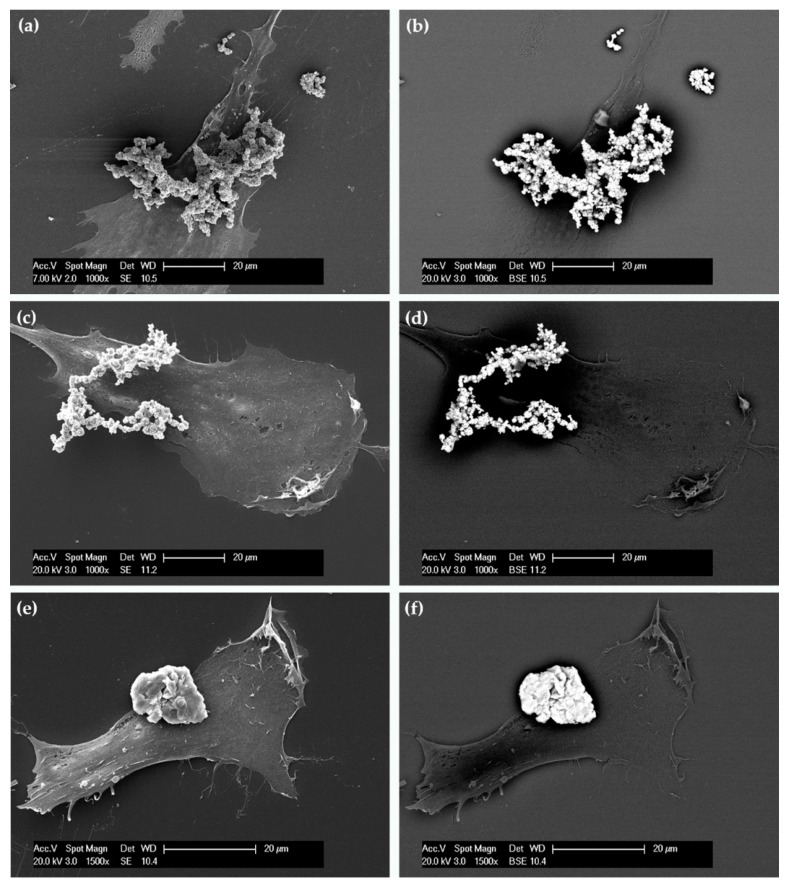
Figure 5.
Scanning electron micrographs of typical hASCs exposed for 24 h to FeMPs (a,b), CoMPs (c,d), and NiMPs (e,f). The same field of view is shown with secondary electron (SE, left) and backscattered electron (BSE, right) imaging. In BSE imaging, the clusters of metal microparticles appear as bright spots.

