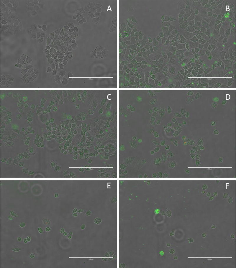
Figure 10.

Images of the Annexin V assay on H460 cells grown in 6-well plates using compound 2 in 10% by weight 2-HPβCD aqueous solution as the compound treatment. All images taken using a 20× objective. Images are presented as a merged image of the normal transmitted light, green fluorescence and red fluorescence figures. (A) 2-HPβCD control, 20 hours. (B) Cisplatin control, 20 hours. (C) Compound 2, 12 hour. (D) Compound 2, 14 hours. (E) Compound 2, 17 hours. (F) Compound 2, 20 hours. Scale bars equal 200 μm.
