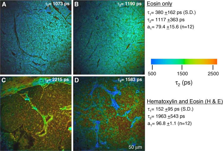
Fig. 2.

Fluorescence lifetime of stained mouse breast tumor samples. a, b In slides stained with eosin only, the fluorescence lifetime is similar throughout the field of view. Regions that encompassed cells and ECM are shown. c, d This value for eosin fluorescence lifetime was preserved in the extracellular matrix regions of H&E stained slides, where hematoxylin does not compete for staining. However, the lifetime of eosin is significantly lengthened in cells compared to eosin only. For each condition, two exemplar images out of 12 are shown. Population averages for the lifetime values are given on the right and the τ2 value for each individual panel shown is indicated
