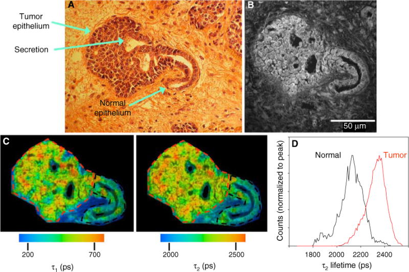
Fig. 4.

The fluorescent properties of normal and tumor epithelium differ. a Digital camera image of H&E stained mouse breast tumor. At this stage of tumor progression, which shows a carcinoma in situ, the distinction between normal epithelium and tumor is evident. Secretion of milk proteins into the remaining duct was preserved in the fixation procedure and, interestingly, was observed to be fluorescent. b Fluorescence intensity image of the sequential unstained slide at 890 nm excitation. c Color maps of the τ1 (left) and τ2 (right) components of the fluorescence lifetime, which illustrate the relatively longer lifetime values in tumor cells when compared to normal epithelium. d Histogram analysis measuring the range of lifetime values of the two ROIs drawn in (c) reveals the shift to longer lifetimes in tumor (red lines) cells compared to normal epithelium (color online)
