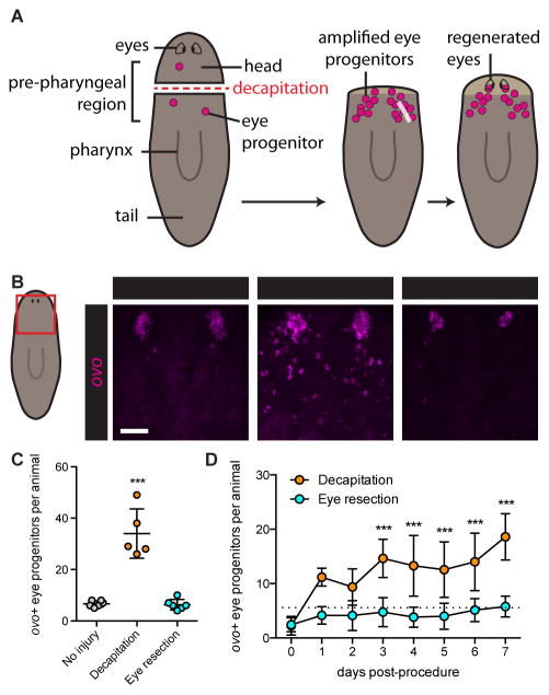
Figure 2. Eye absence is not sufficient to induce eye progenitor amplification.
(A) Schematic of eye regeneration following decapitation. Decapitation (left) leads to amplification of ovo+ eye progenitors (center), which migrate anteriorly and coalesce to form regenerating eyes (right).
(B) Fluorescence in situ hybridization (FISH) with ovo RNA probe, 3 days after indicated surgeries. Maximum intensity projections. Red box on cartoon indicates displayed region. Asterisks mark presumptive eyes, identifiable as coalesced ovo+ cells anterior to progenitors. Scale bar, 50 μm.
(C and D) ovo+ eye progenitor numbers 3 days (C) and 0 to 7 days (D) post-surgery. Data represented as mean ± SD. Dots reflect eye progenitor numbers for individual animals for (C), means for (D). In (D), dotted line reflects mean of uninjured animals on Day 0. Decapitation but not eye resection resulted in significantly elevated eye progenitor numbers in comparison to uninjured controls. n≥4 animals per condition. Statistical significance assessed with respect to uninjured animals by one-way ANOVA (***p<0.001). See also Figures S1D–S1I.

