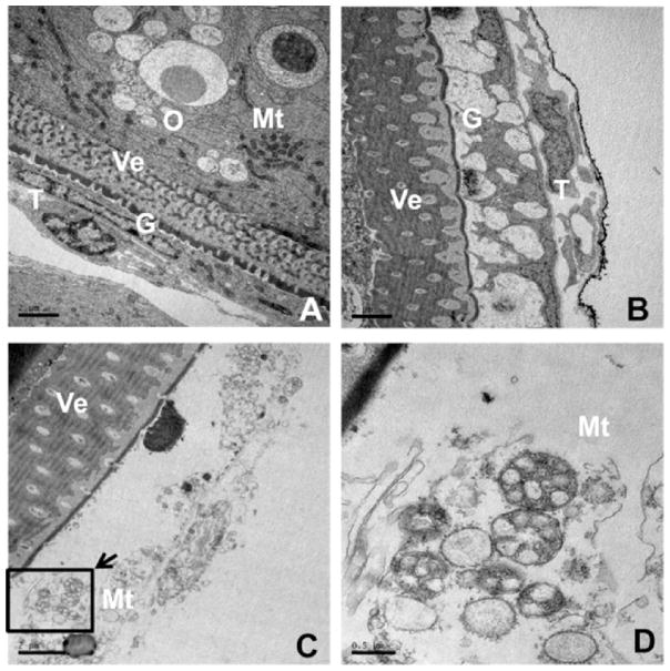
Fig. 4.
Effects of silver nanoparticles (AgNPs) or AgNO3 on ultrastructure of ovarian follicle cells. Ovarian follicles enclosing fully-grown immature oocytes were incubated with Cortland medium containing designed reagents for 2 h. (A), control group showing normal appearance of ovarian follicle cells (Scale bar, 2 μm); (B) AgNPs (30 μg/mL) and (C) AgNO3 (10 μg/mL) treated group showing ovarian follicle cells have irregular cell morphology, acute vacuolation, nuclear condensation and fragmentation (Scale bar, 2 μm); (D) a high magnification image of the area indicated in C showing a novel mitochondrial swelling with intact inner mitochondrial membrane (Scale bar, 0.5 μm). G: granulosa, Mt: mito-chondria, O: oocyte, T: thecal cell, Ve: vitelline envelope.

