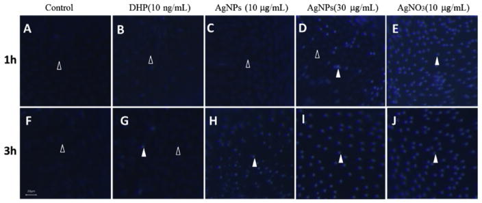
Fig. 5. Effects of silver nanoparticles (AgNPs) or AgNO3 on apoptosis in ovarian follicle cells.
Ovarian follicles enclosing fully-grown immature oocytes were incubated with Cortland medium (control, panel A,F); or medium containing DHP (10 ng/mL, panel B,G); AgNPs (10 μg/mL, panel C,H); AgNPs (30 μg/mL, panel D,I); and AgNO3 (10 μg/mL, panel E,J) that were collected 1 or 3 h following treatment. After Hoechst 33342 staining, the representative fluorescence images were taken to showing the follicle cells layer surrounding the oocytes. Open arrowhead indicated the intact follicle cells with oval shape and their nuclei were stained with a weak blue fluorescence; Filled arrowhead indicated the follicle cells exhibit apoptosis features such as spherical bead shape and bright blue fluorescence when treated with AgNPs or AgNO3. Scale bar, 20 μm.

