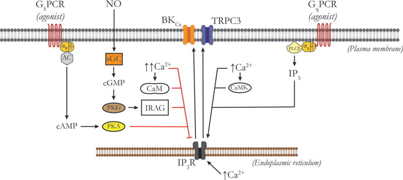
Figure 10.
Regulation of IP3 receptors by vasoconstrictors and vasodilators. Schematic of the plasma membrane and the ER membrane of a vascular SMC showing, from left to right in the plasma membrane, a Gs-protein-coupled receptor, associated G-proteins and AC, a BKCa channel, a TRPC3 channel and a Gq-protein-coupled receptor (GqPCR), associated G-proteins and phospholipase C-β (PLCβ), and an IP3 receptor (IP3R) in the membrane of the ER. Increases in IP3 and moderate increases in cytoplasmic Ca2+ in the environment of an IP3R are the primary stimulae for activation. Activation of IP3Rs by cytosolic Ca2+ is mediated by direct actions of Ca2+ on the channels, or through activation of calcium-calmodulin-dependent protein kinase (CaMK) and phosphorylation of the channels. Vasoconstrictors acting through GqPCRs and activation of PLCβ increase the production of IP3, stimulating Ca2+ release through IP3R. Activated IP3Rs have been shown to physically interact with, and activate plasma membrane BKCa and TRPC3 channels, as shown. Conversely, high levels of intracellular Ca2+ inhibit IP3R activity through mechanisms involving the Ca2+ binding protein, CaM. NO, through activation of soluble guanylate cyclase, increased production of cGMP and activation of PKG phosphorylates IRAG which inhibits IP3R activity. Vasodilators that act at GsPCRs (isoproterenol, adenosine, prostacyclin, CGRP, etc.), activate AC to increase producting of cAMP which then can activate PKA to phosphorylate IP3Rs to decrease their activity. See text for more information.

