
Fig. 1.
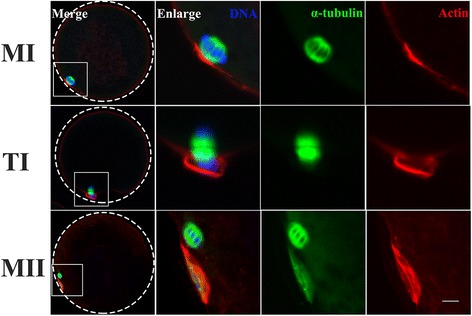
Dynamic distribution of the cytoskeleton during the pig MI-to-MII transition. Samples were taken at MI, TI and MII stages. Microtubules organized bipolar spindles that were symmetrical and barrel in shape, and homologous chromosomes were arranged on the equatorial plate at MI stage; α-tubulin assembled the typical spindle structure and chromosomes were observed around α-tubulin that were in the two polar regions at TI stage. One small polar body was extruded and the microtubules were organized in a bipolar, barrel-shaped structure at the cortex below the polar body, and chromosomes were arranged on the equatorial plate at the cortex in MII stage. Microfilaments frequently formed an actin cap at MI and MII stages and formed a contractile ring at ATI stage. Green, microtubules (α-tubulin); red, microfilaments; blue, chromosomes. Scale bar, 20 μm