Fig. 2.

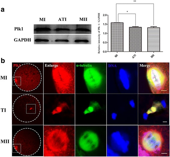
Expression and subcellular localization of Plk1 in porcine oocytes during the MI-to-MII transition. (a) Expression of Plk1 was examined using western blot analysis. Plk1 was expressed in porcine oocytes during the MI-to-MII transition, and a relatively higher Plk1 protein level was detected in MI compared to ATI and MII stages; * P < 0.05; ** P < 0.01. (b) Subcellular localization of Plk1 in porcine oocytes using immunofluorescent staining. Plk1 was enriched at spindle pole regions at MI and MII stages and closely overlapped with α-tubulin leading to the barrel-shaped spindle. Plk1 was associated with the spindle midzone region at TI stage. Red, Plk1; green, spindle; blue, chromosome. Scale bar, 20 μm
