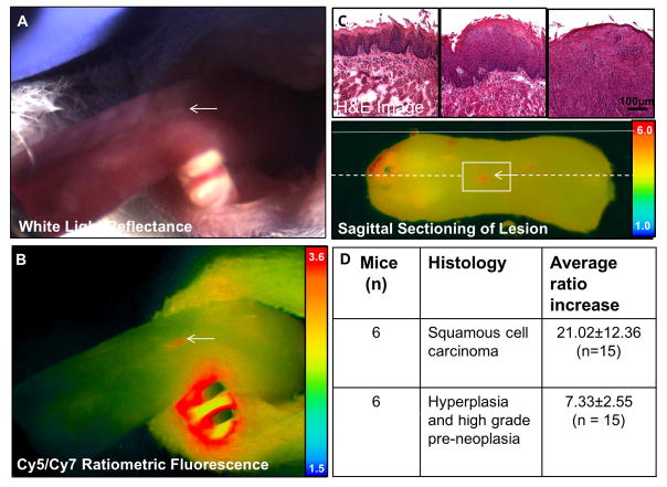
Figure 4. MMP cleavable RACPP detects SCCA not detectable by white light.
A. Dorsal tongue with marked location (white arrow) showing no clear signs on cancer on white light examination B. Ratiometric fluorescence shows increase in Cy5:Cy7 ratio compared to adjacent tissue due to MMP mediated cleavage of RACPP. C. H&E image at the level of boxed region in C showing that the lesion seen in A and B is an invasive carcinoma (third column). There is adjacent low grade and high grade dysplasia in the surrounding tissue (first and second columns). Ex vivo ratiometric fluorescence image showing position on excised tongue and serial sectioning used to confirm pathology of small lesion detected by ratiometric fluorescence. Solid white line shows the position of the lateral tongue edge and stippled white line shows the sagittal section across the lesion. D. Summary table of lesions with corresponding histology that are seen by ratiometric fluorescence but not by white light inspection of the tongue.

