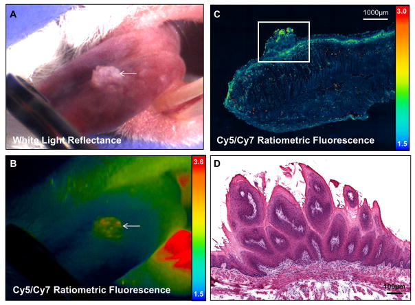
Figure 5. Papilloma detectable by white light reflectance with low RACPP signal.
A. Color image under white light and B. pseudo-color ratiometric fluorescence image showing a large lesion on the dorsal tongue (arrow) in a wild type mouse. C. Confocal image of a sagittal section of tissue across the lesion. There is some evidence of chronic inflammation in the superficial stroma indicated by high fluorescence signal. D. H&E image at the level of boxed region in C showing that the lesion in A and B features benign hyperkeratosis and hyperplasia with finger-like stromal expansion characteristic of papilloma.

