
Fig. 1.
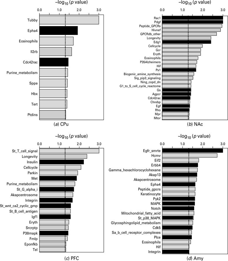
Biological pathways significantly modified by gestational nicotine treatment in the adolescent brain regions analysed by GSEA (nominal p<0.05). For each brain region, the pathways were plotted in descending order of the negative logarithm of their p values at base 10. The biological pathways related to cell adhesion molecules are shown in black columns, whereas others are shown in light grey. For each pathway, a short format of its name in the GSEA database is shown in the figure: for amygdala, Gamma_hexachlorocyclohexane and Mitochondrial_fatty_acid are short formats of Gamma_ hexachlorocyclohexane_degradation and Mitochondrial_fatty_acid_betaoxidation, respectively; for NAc, Sig_pip3_signalling corresponds to Sig_pip3_signalling_in_cardiac_myoctes; for PFC, St_T_cell_signal and St_B_cell_antigen are short formats of St_T_cell_signal_transduction and St_B_cell_antigen_receptor, respectively; for PVN, Oxidative_phosph, Glycerolipid and Sa_B_cell_receptor are short formats of Oxidative_phosphorylation, Glycerolipid_metabolism and Sa_B_cell_receptor_ complexes, respectively; for the other pathways, the word pathway has been omitted from their names.