
Fig 1. Temporary eradication of gut microbiota prevents oxaliplatin-induced mechanical hyperalgesia.
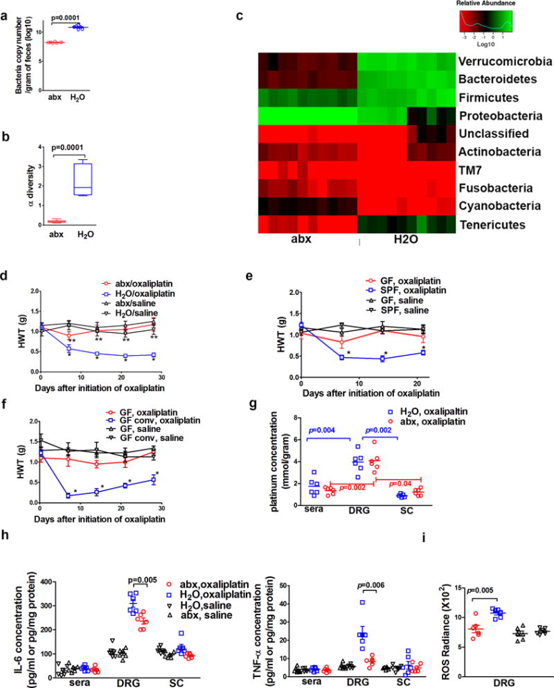
a–c) Impacts of antibiotic water feeding on mice gut microbiota. Fecal samples were obtained after three weeks of antibiotic water (abx, N=10) or regular water (H2O, N=10) followed by DNA isolation from these samples. a) Antibiotics feeding reduced bacterial load as determined by semi-quantitative real-time PCR. b) Antibiotics feeding reduced the α diversity of microbiota. c) Antibiotics feeding altered bacterial community structure as shown in the phylum analysis. d) Gut microbiota eradication prevented the development of oxaliplatin-induced mechanical hyperalgesia. Mice were fed on antibiotics water (abx) or regular water (H2O) prior to oxaliplatin or saline treatment (as control). Hindpaw mechanical withdrawal threshold (HWT) was examined at indicated time points after oxaliplatin therapy. N=6 each group. * p<0.05 H2O/oxaliplatin vs. abx/oxaliplatin. **p>0.05 abx/oxaliplatin vs. abx/saline or H2O/saline. e) Germ-free (GF) status protected mice from oxaliplatin-induced mechanical hyperalgesia. GF or specific pathogen free (SPF) mice were given oxaliplatin or saline. HWT was examined at indicated time points after oxaliplatin therapy. * p<0.05 SPF, oxaliplatin vs. all other groups. N=7 each group. f) GF conventionalization abrogated the protection of mechanical hyperalgesia offered by GF status. To conventionalize GF mice, feces from SPF mice were diluted with PBS and administered daily via gastric gavage for three weeks. GF and conventionalized GF (GF conv) mice were treated with oxaliplatin or saline. Conventionalization of GF mice abrogated the protection offered by GF status. * p<0.05 GF conv, oxaliplatin vs. GF, oxaliplatin. N=8 for GF, oxaliplatin; N=7 for GF, saline; N=7 for GF conv, oxaliplatin; N=6 for GF conv, saline. g) Gut microbiota eradication did not change tissue oxaliplatin distribution (N=6 each group). Spinal cord (sc), serum and dorsal root ganglion (DRG) concentrations of platinum were determined by ICP-MS. Two-way ANOVA test suggested abx treatment did not change tissue platinum distribution (p=0.42) while types of tissues have significant influence on platinum levels (p=0.001). Post-hoc t-tests (p values in the figure). were performed to identify the impact of tissue type on platinum levels h) DRG cytokine levels for IL-6 and TNF-α were lower in mice with eradicated gut microbiota than those in mice with normal gut microbiota (N=6 each group, one-way ANOVA indicated there was significant difference in DRG samples, and subsequent post-hoc t-test was used to determine the difference between the abx and H2O groups). Sera and spinal cord levels for IL-6 and TNF-α were not significantly different among groups (N=6 each group, one-way ANOVA test for sera IL-6 samples, p=0.58; spinal cord IL-6 samples, p=0.06; sera TNF-α samples, p=0.88; spinal cord TNF-α samples, p=0.75). i) Reduced levels of reactive oxygen species (ROS) in DRG from mice with eradicated gut microbiota. At day 10 after the initiation of oxaliplatin therapy, levels of ROS were determined with IVIS using L-012 as a chemiluminescent probe. N=6 each group. One-way ANOVA test followed by post-hoc test were used for statistical analysis.