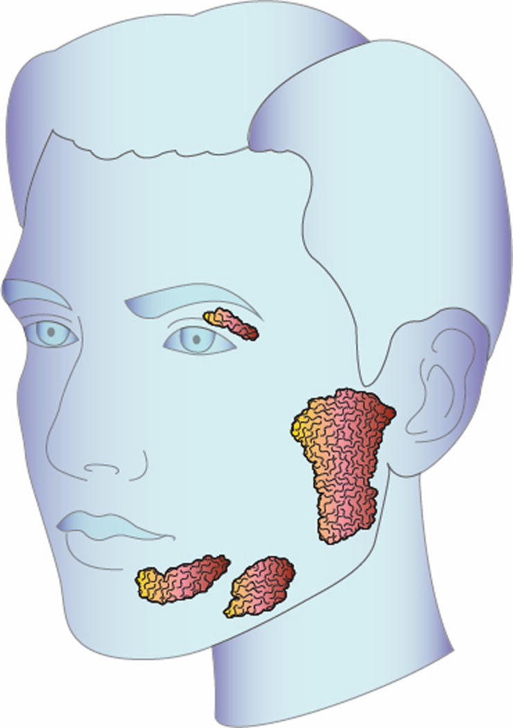
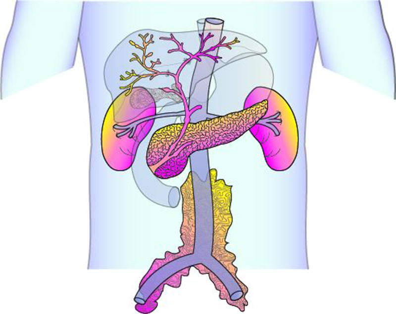
Figure 1.


(A) Head and neck illustration highlighting involvement of the lacrimal and major salivary glands. Lacrimal and salivary gland enlargement is most often bilateral in distribution. (B) Abdomen illustration highlighting typical organ involvement including the pancreas, bile ducts, kidneys, and retroperitoneal tissue. Radiographically, the retroperitoneal fibrosis often extends inferiorly to encase the iliac vessels. Not depicted here, the aorta and lung are other common sites of disease involvement.
