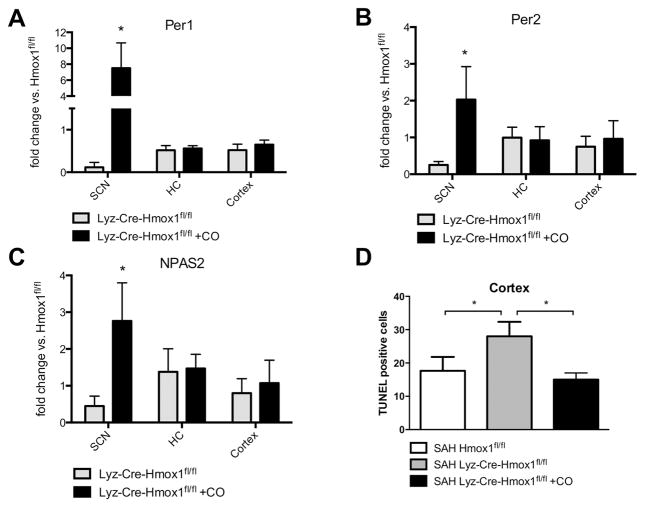
Figure 6. Myeloid HO-1 and carbon monoxide (CO) modulate circadian rhythm gene expression and susceptibility to neuronal injury.
A–D. Expression of Per-1 (A), Per-2 (B) and NPAS-2 (C) in the indicated brain regions (SCN, HC, cortex) in Lyz-Cre-Hmox1fl/fl mice ± CO treatment for 7 days after SAH compared to Hmox1fl/fl control animals. Lyz-Cre-Hmox1fl/fl vs. Lyz-Cre-Hmox1fl/fl + CO, change in the SCN: *p=0.0156 for Per-1 (A); *p=0.0267 for Per-2 (B); *p=0.0201 for NPAS-2 (C). Results represent mean ± SD from 3–4 mice/treatment group. D. Quantification of TUNEL-positive cells per microscopic field in the cortex of Hmox1fl/fl and Lyz-Cre-Hmox1fl/fl mice ±CO: Hmox1fl/fl vs. Lyz-Cre-Hmox1fl/fl *p=0.0408; Lyz-Cre- Hmox1fl/fl vs. Lyz-Cre-Hmox1fl/fl +CO *p=0.0146. Results represent mean ± SD of 3–4 mice/group.

