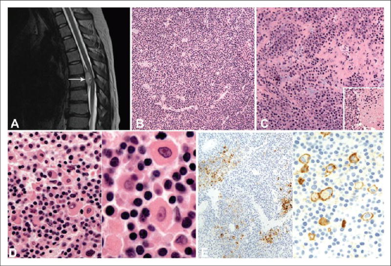
Figure 1.
(a), MRI without contrast shows the T5–T6 ventral epidural tumor (arrow) that measured 2.4 cm and is T2 hyperintense, but not isointense with cerebrospinal fluid. A small (9 mm) fluid-containing focus within the tumor is radiologically consistent with cystic change. (b), Hematoxylin and eosin (H&E) section shows a diffuse arrangement of neoplastic cells with prominent vasculature. (c), The neoplastic cells are focally noted in a fibrous and myxoid stroma. Inset: Focal necrosis is noted within the tumor. (d), On high-power magnification, a large proportion of cells demonstrate moderate cytoplasm and nuclei with coarse chromatin (left and right panels). A smaller proportion of neoplastic cells demonstrate more abundant eosinophilic cytoplasm including cells with rhabdoid or plasmacytoid morphology (left panel). Occasional cells have nuclei with vesicular chromatin and prominent nucleoli (right panel). (e), An immunohistochemical stain with the CD99 antibody. Low-power magnification demonstrates membranous staining of a small proportion of neoplastic cells (left panel). On high-power magnification, membranous staining of rhabdoid/plasmacytoid cells is noted (right panel).

