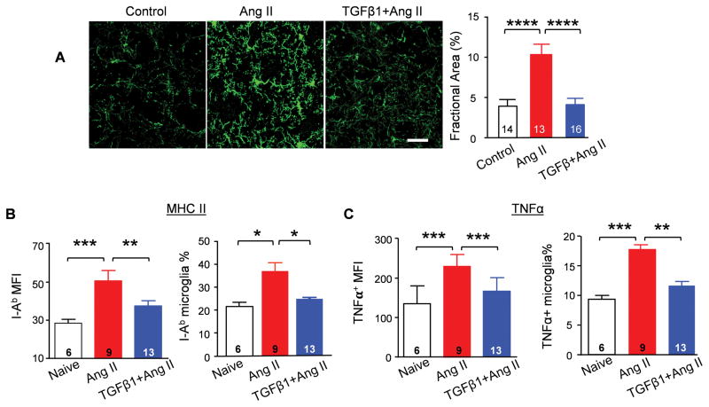
Figure 3. TGFβ1 suppresses microglial activation.
(A) Representative immunostaining of microglia with anti-Iba1 antibody in the PVN. Calibration bar equals to 20 μm. Fractional area of Iba-1 staining was analyzed in each group. **** P<0.0001. Expression of MHC class II subtype I-Ab (B) and TNFα (C) in CD11b+CD45low-microglia was evaluated by flow cytometry analysis. *P<0.05, **P<0.01, ***P<0.001.

