
Figure 6.
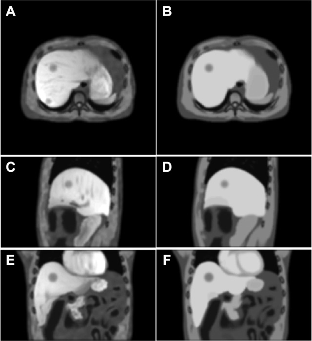
A comparison of the end of exhalation (EOE) volume between the developed magnetic resonance (MR) phantom (left column) and the pseudo-MR volume generated by the current extended cardiac-torso (XCAT) phantom with uniform intensity assignment (right column) at axial (A and B), sagittal (C and D), and coronal (E and F) views.