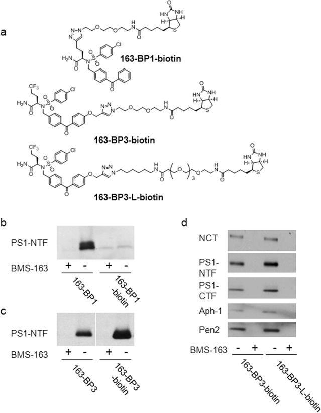
Figure 3.

Biotinylated photoaffinity probes. (a) Chemical structure of the biotinylated probes. (b, c) Biotinylated probes and clickable probes (20 nM) were incubated with HeLa membranes in the presence or absence of 1 μM competitor BMS-163 and UV-irradiated, followed by click chemistry with biotin azide for clickable probes only, pull-down with streptavidin resin, and Western blot analysis for PS1–NTF. (d) The solubilized γ-secretase was captured under native conditions (0.25% CHAPSO) by 163-BP3–biotin and 163-BP3–L-biotin. The bound complex was eluted and analyzed by Western blot analysis.
