Fig. 2.
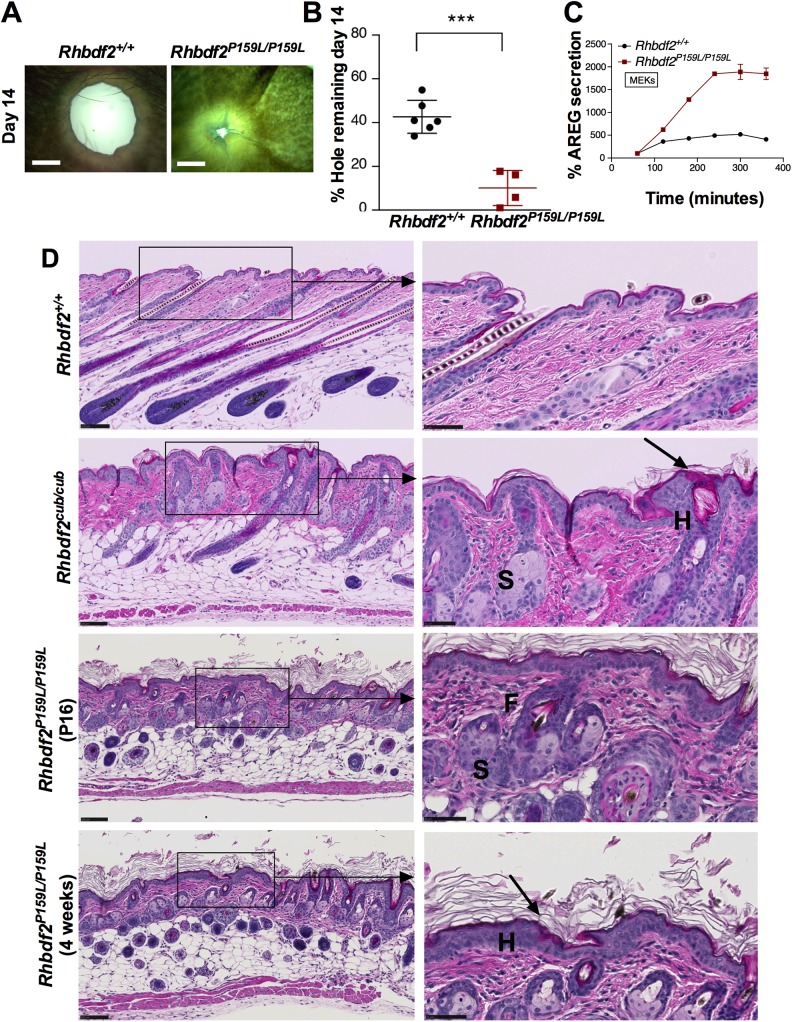
Rapid wound healing and proliferative skin phenotype in Rhbdf2P159L/P159L mice. (A) Representative images of regenerating ear tissue in eight-week-old female Rhbdf2+/+ and Rhbdf2P159L/ P159L mice at 14 days postwounding. Magnification, 4×; scale bars: 1 mm. (B) Quantification of ear hole closures shown in A. Rhbdf2+/+ (n=6) and Rhbdf2P159L/P159L (n=4). (C) ELISA quantitation of AREG levels in the supernatants of cultured Rhbdf2+/+ and Rhbdf2P159L/P159L MEKs in response to stimulation with 100 nM PMA for the indicated times. (D) H&E-stained skin sections of female Rhbdf2+/+, Rhbdf2cub/cub and Rhbdf2P159L/P159L mice. Both the Rhbdf2cub/cub and Rhbdf2P159L/P159L mice present with significant hyperplasia (H), hyperkeratosis (arrow), enlarged sebaceous glands (S), and follicular dystrophy (F) compared with the Rhbdf2+/+ mice. Scale bars: 100 μm (low magnification); 50 μm (high magnification).

