Fig. 1.
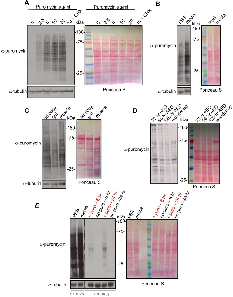
Puromycin labelling to measure protein synthesis during larval development. (A) Whole inverted third instar larvae were incubated in increasing amounts of puromycin (5 µg/ml), or with puromycin (5 µg/ml)+cycloheximide (CHX, last lane) together, for 40 min. Equal amounts of whole larval protein extracts were then analyzed by western blotting. Left, western blot with either anti-puromycin, or anti-tubulin antibodies. Right, Ponceau S staining showing total protein levels. (B) Whole inverted larvae were incubated in either PBS+puromycin (5 µg/ml) or Schneider's media+puromycin (5 µg/ml) for 40 min. Equal amounts of whole larval protein extracts were then analyzed by western blotting. Left, western blot with either anti-puromycin, or anti-tubulin antibodies. Right, Ponceau S staining showing total protein levels. (C) Whole inverted third instar larvae were incubated in Schneider's media+puromycin (5 µg/ml) for 40 mins. Larval tissues were then isolated and analyzed by western blotting. Left, western blot with either anti-puromycin, or anti-tubulin antibodies. Right, Ponceau S staining showing total protein levels. (D) Larvae at different stages in development (72 h AED, 96 h AED, 120 h AED and wandering stage) were inverted and incubated in Schneider's media+puromycin (5 µg/ml) for 40 min. Equal amounts of whole larval protein extracts were then analyzed by western blotting. Left, western blot with anti-puromycin. Right, Ponceau S staining showing total protein levels. (E) Comparing ex vivo versus in vivo feeding for puromycin labelling. For the ex vivo experiments, third instar larvae were inverted and incubated in either PBS+puromycin (5 µg/ml) or Schneider's media+puromycin (5 µg/ml) for 40 min. For the feeding experiments, third instar larvae were transferred to either normal food (no puro) or normal food supplemented with 25 µg/ml of puromycin (+ puro) for either 6 or 24 h. For both the ex vivo and in vivo samples, equal amounts of whole larval protein extracts were then analyzed by western blotting. Left, western blot with either anti-puromycin, or anti-tubulin antibodies. Right, Ponceau S staining showing total protein levels. Note, the vertical dotted line in the western blots indicates where the blot was spliced to remove an empty lane and the molecular weight ladder lane (see Ponceau S staining). All experiments were carried out using w1118 larvae.

