
Figure 4.
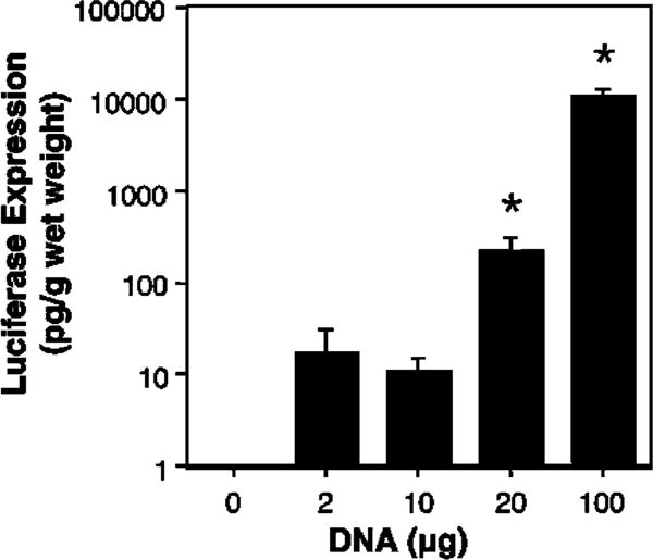
Dose curve for electroporation-mediated gene transfer. Mice were injected intratracheally with varying concentrations of pCMV-Lux-DTS (200 μl) and electroporated at 200 V/cm as described in Figure 2 (n = 4 animals per concentration). After 2 days, luciferase activities were measured as described in the section materials and methods. Mann–Whitney U-tests were performed to determine statistical significance. *P<0.001 compared to no DNA.