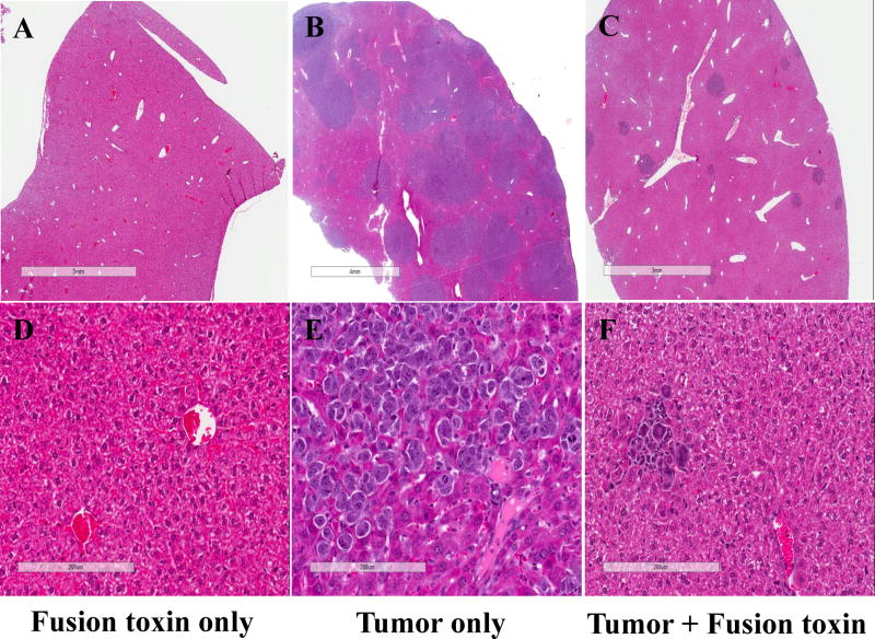
Fig. 4.
Pathology analysis: A and D) Liver from an NSG mouse injected with bivalent human IL-2 fusion toxin at 100 μg/kg shows unremarkable hepatic parenchyma with no increase in hepatocyte necrosis or inflammation. B and E) Liver from a mouse injected with HUT102/6TG tumor cells shows replacement of liver parenchyma by sheets of malignant cells with condensed chromatin, prominent nucleoli, an elevated nuclear: cytoplasmic ratio, and geographic necrosis. C and F) Liver from a mouse injected with both HUT102/6TG tumor cells and bivalent human IL-2 fusion toxin at 100 μg/kg shows normal hepatic parenchyma in most of the examined section and a few sporadic malignant cells.

