Figure 2.
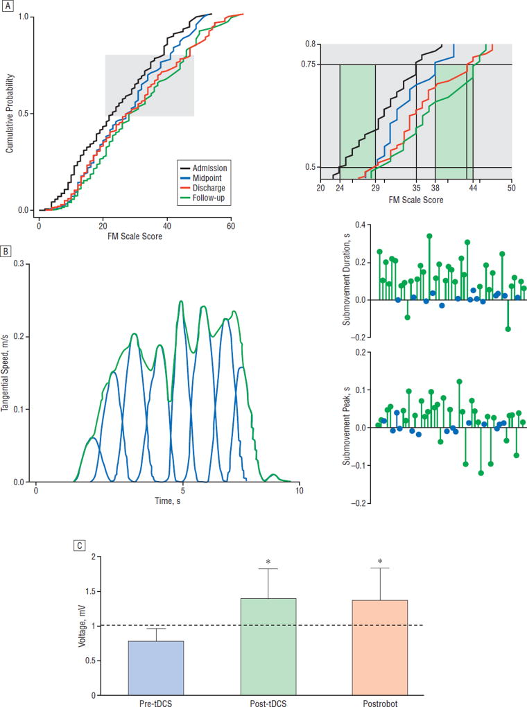
Results from treatments using robotic devices. A, Cumulative probability distributions of total Fugl-Meyer Motor (FM) scale scores (range, most severe=0 to less severe=54; maximum, 66) for patients evaluated at treatment admission, midpoint, treatment discharge, and follow-up 3 months after robotic training has ended. The curve representing this group of treated patients shifts to higher FM scores at the midpoint of training and it shifts to the right at discharge and follow-up indicating progressive increase of motor ability as the FM scores increase. The improvement after training is significant (Kolmogorov-Smirnov test, P<.001). Closer inspection of the cumulative probability values between 0.5 and 0.75 (right side of part A) are taken from the gray region depicted in the main graph. At the 0.5 level, admission to midpoint captures most of the improvement. At the 0.75 level, the improvement occurs after the midpoint evaluation and again at follow-up. For some patients with less severe impairment, even an intensive training experience did not define a performance optimum, as there was additional improvement after discharge. For other patients, the dose of training needs to be optimized. B, The left panel demonstrates the speed with which the patient executes an untrained movement at the start of training. The movement can be decomposed into submovements. The right panels depict an analysis of the individual submovements and, in particular, the peak speed and the duration of each submovement. A patient’s performance on this untrained task is represented by a line from the baseline performance connected to a green (P<.05) or a blue circle (P>.05), and each circle represents the discharge value. At discharge, most patients demonstrate longer submovements that are executed more quickly. These changes in submovements reflect smoother movement.10 C, Transcranial magnetic stimulation generates motor evoked potentials (MEPs) in the flexor carpi radialis (N=6 patients, bars represent mean [SEM]). The MEPs are recorded from the flexor carpi radialis muscle during a low-level isometric wrist flexion, before and immediately following 20-minute anodal transcranial direct current stimulation (tDCS), then again after 1 hour of robotic wrist therapy. Following tDCS, MEP amplitude is significantly elevated (*P<.05) and remains significantly elevated after robotic therapy (*P<.05), indicating integrity and potentially increased efficiency within the corticomotor pathways.33

