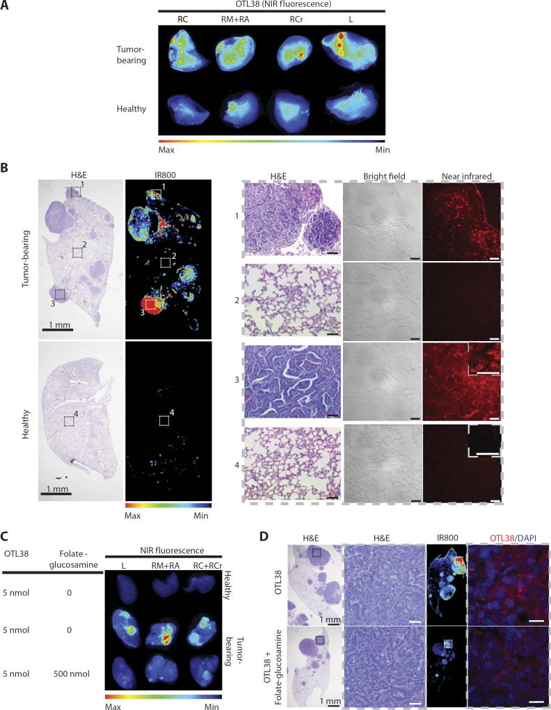
Fig. 4. Murine KrasLSL-G12D/+;p53flx/flx lung adenocarcinomas express FR.
(A) NIR fluorescence imaging showing that ligand OTL38 (On Target Laboratories), an FRα-targeting ligand conjugated to a NIR dye, is preferentially retained in lung tumors and cleared from normal healthy lungs. KrasLSL-G12D/+;p53Flox/Flox mice were injected with 5 nmol of OTL38 8 weeks after tumor induction and sacrificed 24 hours after injection. Whole lungs were excised and imaged using LI-COR Odyssey CLx. A noninduced healthy mouse was used as a control. RC, right caudal lobe; RM, right medial lobe; RA, right accessory lobe; RCr, right cranial lobe; L, left lobe. (B) Histological and NIR images of right lung lobes from mice treated with OTL38. Left: Whole-organ low-magnification hematoxylin and eosin (H&E) and corresponding NIR images. Right: High-magnification H&E, bright-field, and NIR images of tumorous and healthy tissue corresponding to insets in (B). Scale bars, 50 µmand20 µm (inset). (C) Whole-organ NIR fluorescence images of excised lung lobes from mice bearing lung tumors treated with OTL38 (5 nmol) in the presence or absence of ≥100-foldmolar excess of folate-glucosamine (n = 3 per group). (D) Representative H&E and fluorescence images of tissues from (C). Insets on low-magnification H&E images correspond to adjacent high-magnification images of tumor tissue. Insets on whole-organ NIR fluorescence images correspond to adjacent high-magnification NIR images. H&E images represent the types of tissue shown in NIR images. Scale bars, 20 µm.

