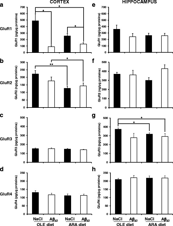
Fig. 4.

Modification of α-amino-3-hydroxy-5-methyl-4-isoxazolepropionic acid (AMPA) receptors in cortex and hippocampus homogenates induced by arachidonic acid-enriched (ARA) diet and intracerebroventricular injections of amyloid-β peptide 42 oligomers. Immediately after the probe test, mice were killed, and homogenates were prepared from cortex and hippocampus. The expression levels of the four murine members of the AMPA receptor family were measured in cortex (a–d) and hippocampus e–h homogenates by using specific enzyme-linked immunosorbent assay (ELISA) kits from Aviva Systems Biology Corporation for glutamate receptor 1 (GluR1) (a and e) and GluR2 (b and f) and from Cloud-Clone Corporation for GluR3 (c and g) and GluR4 (d and h). Data are expressed as picograms of the specific AMPA receptor family member per microgram of total protein in the brain homogenates (* p < 0.05 and ** p < 0.01, comparing the four groups of mice). Results are shown as mean ± SEM of ELISA measurements performed for all animals of the group (oleic acid-enriched diet [OLE] groups, n = 4; ARA groups, n = 6). Measurements were performed in duplicate for each brain tissue sample
