
Fig. 6.
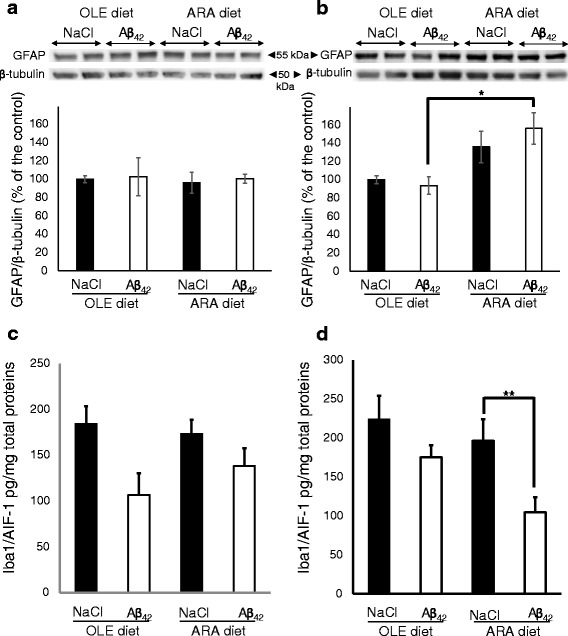
Modifications of brain glial fibrillary acidic protein (GFAP) and Iba1/AIF-1 proteins induced by arachidonic acid-enriched (ARA) diet and amyloid-β peptide 42 (Aβ42). Immediately after the probe test, mice were killed, and homogenates were prepared from the cortex and hippocampus. Representative immunoblots of cortical (a) and hippocampal (b) GFAP astroglial protein from oleic acid-enriched (OLE) or ARA diet mice after NaCl or Aβ42 injections are shown. Densitometric analyses were performed to determine signal intensities normalized to β-tubulin, and data are expressed as percentages of control OLE mice injected with NaCl (* p < 0.05 and ** p <0.01, comparing the four groups of mice). Results are shown as mean ± SEM of immunoblots performed for all animals (OLE groups, n = 4; ARA groups, n = 6). Expression levels of the Iba1/AIF-1 microglial marker was determined by enzyme-linked immunosorbent assay (ELISA), and the relative amounts of Iba1/AIF-1 per microgram of total protein are shown in the cortex (c) and hippocampus (d) of the OLE- and ARA-fed mice after intracerebroventricular NaCl or Aβ42 injections. Results are shown as mean ± SEM of ELISA measurements in each group of mice (OLE groups, n = 4; ARA groups, n = 6)