Fig. 4.

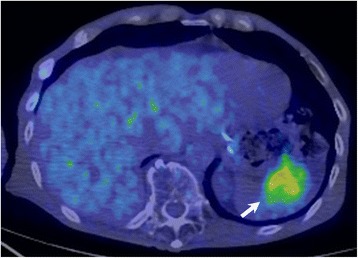
18F-2-deoxy-2-fluoro-glucose (FDG) positron emission tomography combined with computed tomography imaging showing the splenic mass with intense FDG uptake (arrow)

18F-2-deoxy-2-fluoro-glucose (FDG) positron emission tomography combined with computed tomography imaging showing the splenic mass with intense FDG uptake (arrow)